Neuroscience and Neuro-Urology Basic Research
project — From sensory signal to motor action
The brain coordinates sensory input from the lower spinal cord and pelvis, and information from the external environment to ‘decide’ when to void, by activating or inhibiting neurons in the pontine micturition center. We aim to identify the specific neurons that comprise i) the bladder-afferent-activity sensing circuits and ii) the continence-regulating neural circuits; both of which function critically within the reflex micturition pathway.
The lab uses in vivo fiber photometry (6,7) to reveal the temporal sequence underlying PMC neuron activation. Furthermore, we use techniques for manipulating neural activity (8-12), to connect neurons in specific ‘nodes’ of the micturition reflex pathway with roles in spontaneous voiding.
References:
6) Lin et al., (2016) Nat Neurosci. 19, 1142-1153. 7) Chen et al., (2013) Nature 499, 295- 300. 8) Deisseroth. (2015) Nat Neurosci. 18, 1213-1225. 9) Atasoy and Sternson. (2018) Physiol Rev 98, 391-418. 10) Roth. (2016) Neuron 89, 683-694. 11) Anaclet et al., (2018) J. Neurosci. 38, 5168-5181. 12) Todd et al., (2018) Nat Neurosci. 21, 717–724.
Read more:

Schematic of ChR2.mCherry construct and viral targeting unilateral to a PMC-afferent brain site.
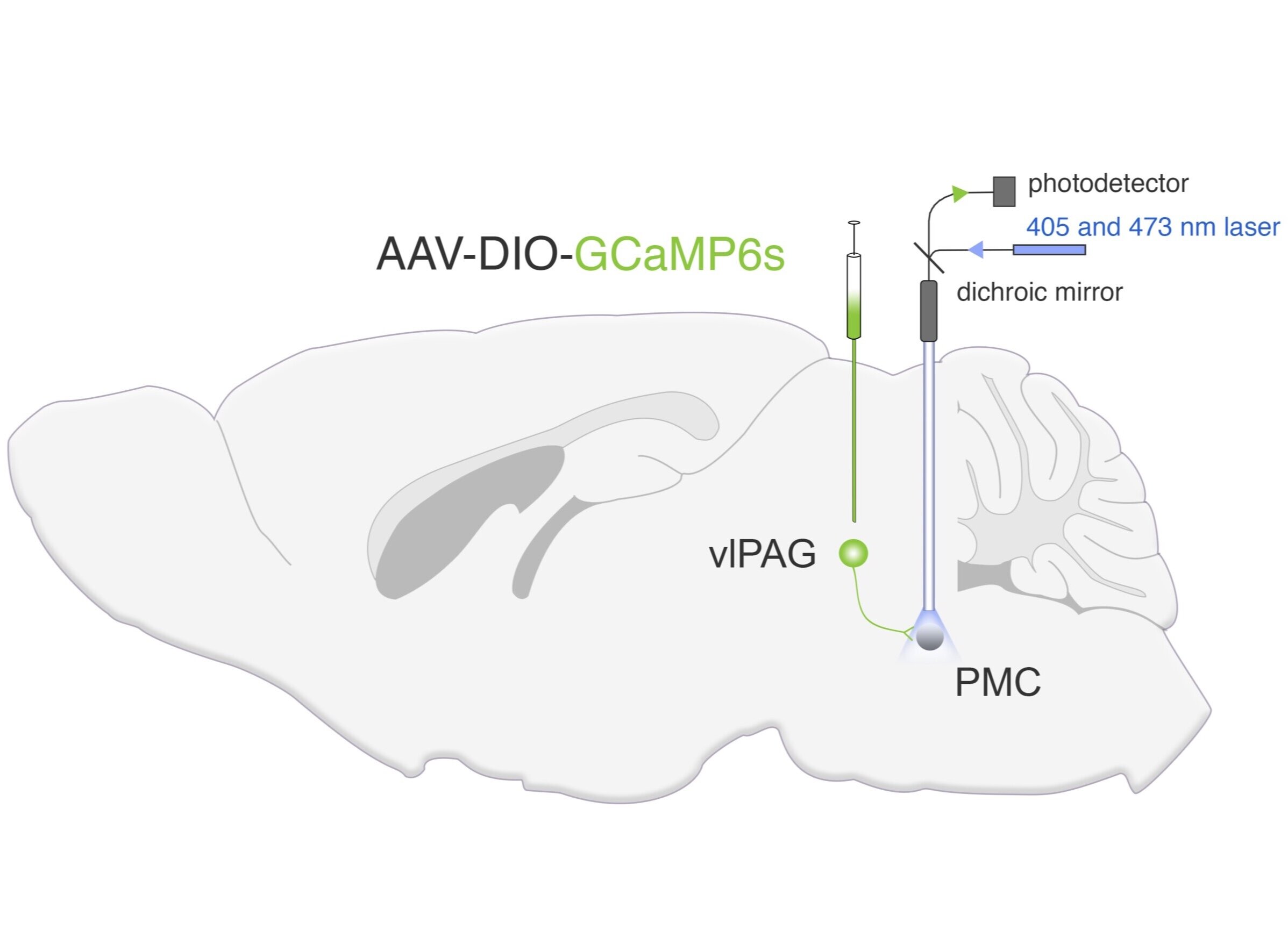
Schematic of GCaMP6s construct and viral targeting unilateral to a PMC-afferent brain site.